Abstract
Foliar trichome morphology, trichome basal insertion cells (TBIC) and cells surrounding trichome insertion basal cells (CSTIBC) on the leaves of fourteen species of family Asteraceae were investigated by light microscopy. TBIC and CSTIBC are the two novel characters investigated in this work, in search of unique character states that could improve the taxonomy of the family. Epidermal peels were made following standard procedure. Data obtained showed that both non-glandular and glandular trichomes were present, the glandular was observed in the genus Vernonia only. The non-glandular trichomes were unicellular, bicellular, tricellular and multicellular uniseriate. Shriveled cells within the multicellular uniseriate cells were common in Emilia sonchifolia and Emilia praertermissa and occurred sparsely in Ageratum conyzoides. Uniquely pigmented multicellular uniseriate trichomes with apical cells shriveled or transparent delimits Bidens pilosa from the other species studied. Amoeboid shaped multicellular uniseriate trichomes delimit Chromolaena odorata from the other species, while Tshaped trichomes (regular and irregular) delimit the genus Vernonia from the other genera studied. Shape and width of apical cells of the trichomes differentiate Vernonia cinerea from Vernonia amygdalina. Surface of trichomes in Aspilia africana, Synedrella nodiflora and Eclipta alba are uniquely papillate and diagnostic for the three species. TIBC have varying shapes with different combinations of 1-4 shapes per species. It is noteworthy that the two species of Emilia can be delimited from each other based on the shapes of the TIBC on the abaxial epidermis. The number of CSTIBC was observed to be the same on the adaxial epidermis of the two species of genus Emilia. Shapes of CSTIBC in the species studied are diverse. Coefficient of Variation for all the characters studied showed species with more variation within each character. Novel characters, TIBC and CSTIBC, were found to be useful as additional tools in the taxonomy of family Asteraceae.
Abstract from the paper’s OpenAlex record, shown for indexing and research. Copyright remains with the authors or the publisher. Read at the source ↗
The text the model read, with the source record’s inline markup (italics, sub- and superscripts) rendered and its character entities decoded; quote offsets refer to the raw text.
Claims 17
Claim · model-written summary
Both glandular and non-glandular trichomes were found in Asteraceae, with glandular trichomes restricted to Vernonia.
- Outcome
- glandulare0
 Vernonia
Organisms pictured · Representative pictures of the matched species, not figures from the paper.

Verbatim
e0“both non-glandular and glandular trichomes were present, the glandular was observed in the genus Vernonia only.”
Claim · model-written summary
Non-glandular trichomes had one, two, three or multiple cells.
- Outcome
- non-glandular trichomese0
Not reportedTaxon
Verbatim
e0“The non-glandular trichomes were unicellular, bicellular, tricellular and multicellular uniseriate.”
Claim · model-written summary
Shriveled multicellular trichome cells were common in Emilia sonchifolia.
- Taxon
- Emilia sonchifolia
 Emilia sonchifoliae0
- Outcome
- Shriveled cellse0
Organisms pictured · Representative pictures of the matched species, not figures from the paper.

Verbatim
e0“Shriveled cells within the multicellular uniseriate cells were common in Emilia sonchifolia and Emilia praertermissa”
Claim · model-written summary
Shriveled multicellular trichome cells were common in Emilia praertermissa.
Verbatim
e0“Shriveled cells within the multicellular uniseriate cells were common in Emilia sonchifolia and Emilia praertermissa”
Claim · model-written summary
Shriveled multicellular trichome cells were sparse in Ageratum conyzoides.
- Taxon
- Ageratum conyzoides
 Ageratum conyzoidese0
- Outcome
- occurred sparselye0
Organisms pictured · Representative pictures of the matched species, not figures from the paper.

Verbatim
e0“occurred sparsely in Ageratum conyzoides.”
Claim · model-written summary
Pigmented uniseriate trichomes with shriveled or transparent apical cells distinguished Bidens pilosa.
- Taxon
- Bidens pilosa
 Bidens pilosae0
- Outcome
- pigmented multicellular uniseriate trichomese0
Organisms pictured · Representative pictures of the matched species, not figures from the paper.

Verbatim
e0“Uniquely pigmented multicellular uniseriate trichomes with apical cells shriveled or transparent delimits Bidens pilosa from the other species studied.”
Claim · model-written summary
Amoeboid uniseriate trichomes distinguished Chromolaena odorata.
- Taxon
- Chromolaena odorata
 Chromolaena odoratae0
- Outcome
- Amoeboid shaped multicellular uniseriate trichomese0
Organisms pictured · Representative pictures of the matched species, not figures from the paper.

Verbatim
e0“Amoeboid shaped multicellular uniseriate trichomes delimit Chromolaena odorata from the other species”
Claim · model-written summary
T-shaped trichomes distinguished Vernonia.
- Outcome
- Tshaped trichomese0
Organisms pictured · Representative pictures of the matched species, not figures from the paper.
Verbatim
e0“Tshaped trichomes (regular and irregular) delimit the genus Vernonia from the other genera studied.”
Claim · model-written summary
Apical trichome cell shape and width distinguish Vernonia cinerea and V. amygdalina.
- Taxon
- Vernonia cinerea
 Cyanthillium cinereume0
- Outcome
- Shape and width of apical cellse0
- Comparator
- Vernonia amygdalinae0
Organisms pictured · Representative pictures of the matched species, not figures from the paper.

Verbatim
e0“Shape and width of apical cells of the trichomes differentiate Vernonia cinerea from Vernonia amygdalina.”
Claim · model-written summary
Aspilia africana has diagnostic papillate trichomes.
- Taxon
- Aspilia africana
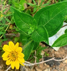 Aspilia africanae0
- Outcome
- Surface of trichomese0
Organisms pictured · Representative pictures of the matched species, not figures from the paper.

Verbatim
e0“Surface of trichomes in Aspilia africana, Synedrella nodiflora and Eclipta alba are uniquely papillate and diagnostic for the three species.”
Claim · model-written summary
Synedrella nodiflora has diagnostic papillate trichomes.
- Taxon
- Synedrella nodiflora
 Synedrella nodiflorae0
- Outcome
- Surface of trichomese0
Organisms pictured · Representative pictures of the matched species, not figures from the paper.

Verbatim
e0“Surface of trichomes in Aspilia africana, Synedrella nodiflora and Eclipta alba are uniquely papillate and diagnostic for the three species.”
Claim · model-written summary
Eclipta alba has diagnostic papillate trichomes.
- Taxon
- Eclipta alba
 Eclipta albae0
- Outcome
- Surface of trichomese0
Organisms pictured · Representative pictures of the matched species, not figures from the paper.

Verbatim
e0“Surface of trichomes in Aspilia africana, Synedrella nodiflora and Eclipta alba are uniquely papillate and diagnostic for the three species.”
Claim · model-written summary
Basal insertion cells had one to four shapes per species.
Not reportedTaxon
Verbatim
e0“TIBC have varying shapes with different combinations of 1-4 shapes per species.”
Claim · model-written summary
The two Emilia species could be distinguished by abaxial basal insertion cell shapes.
- Outcome
- shapes of the TIBCe0
 Emilia
Organisms pictured · Representative pictures of the matched species, not figures from the paper.

Verbatim
e0“the two species of Emilia can be delimited from each other based on the shapes of the TIBC on the abaxial epidermis.”
Claim · model-written summary
The two Emilia species had the same number of surrounding cells on their adaxial epidermis.
- Outcome
- number of CSTIBCe0
Organisms pictured · Representative pictures of the matched species, not figures from the paper.
Verbatim
e0“The number of CSTIBC was observed to be the same on the adaxial epidermis of the two species of genus Emilia.”
Claim · model-written summary
Surrounding cell shapes varied among the studied species.
- Outcome
- Shapes of CSTIBCe0
Not reportedTaxon
Verbatim
e0“Shapes of CSTIBC in the species studied are diverse.”
Claim · model-written summary
The authors found basal insertion and surrounding cells useful for Asteraceae taxonomy.
- Taxon
- AsteraceaeAsteraceaee0
- Outcome
- useful as additional toolse0
- Comparator
- Novel characterse0
Not reportedValue
Verbatim
e0“Novel characters, TIBC and CSTIBC, were found to be useful as additional tools in the taxonomy of family Asteraceae.”